Evaluating the aortic valve is a routine part of an echocardiogram. There are many methods we use to determine the structure and function of the valve, including 2D, color Doppler and a combination of pulsed & continuous-wave Doppler. With short examination times being a common challenge, it can easily cause measurement errors within our studies. This blog is going to cover 4 errors to avoid when measuring continuous-wave (CW) Doppler tracings of the peak aortic valve velocity.
One of the most common errors to properly evaluating the peak aortic valve (AV) velocity, is not having the Doppler cursor parallel to flow. Let’s recall back to the days of learning physics:

Avoid measuring fine linear signals at peak velocity waveform. These fine linear signals should not be included in the velocity tracing.

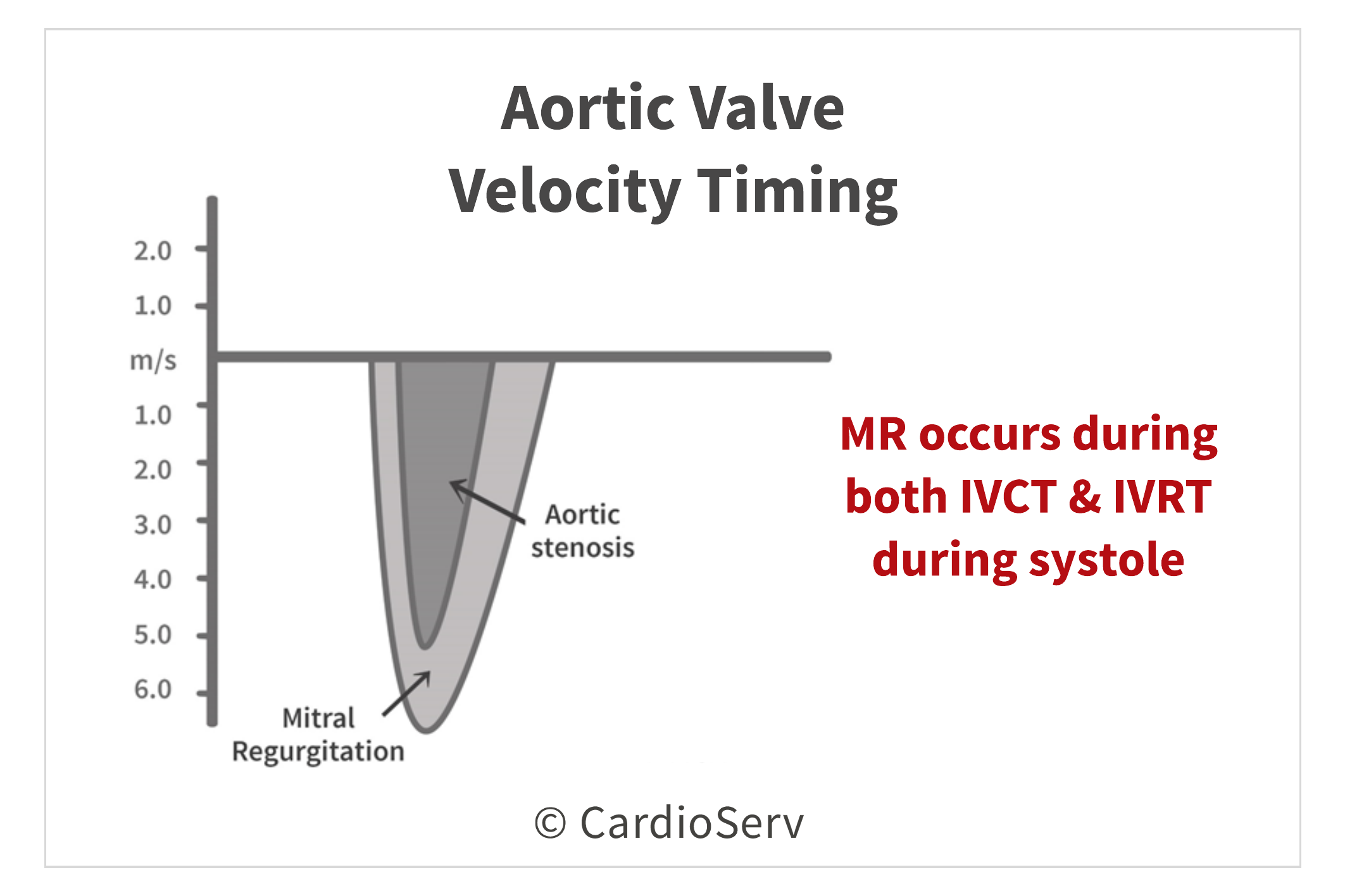
Mitral regurgitation (MR) velocity can easily be mistaken for the aortic valve (AV) velocity if not paying close attention! We are able to differentiate the two by evaluating the timing at which the velocity occurs.


It’s not uncommon for our patients to have PVC’s throughout their exam. Although we cannot prevent this from occurring, we can be sure to not measure these beats! This includes not measuring the extra beat (PVC) and the following velocity jet.

When evaluating the peak aortic valve CW Doppler velocities, be sure to avoid these 4 common errors:

Andrea Fields MHA, RDCS
Stay Connected: LinkedIn, Facebook, Twitter, Instagram
References:
Lang, R. M., MD, Badano, L. P., MD, & Mor-Avi, V., PhD. (2015). Recommendations for Cardiac Chamber Quantification by Echocardiography in Adults: An Update from the American Society of Echocardiography and the European Association of Cardiovascular Imaging. JASE, 28(1), 1-53. Retrieved March 1, 2017, from http://asecho.org/wordpress/wp-content/uploads/2015/01/ChamberQuantification2015.pdf


Feb
2018
Feb
2018
Feb
2018
Feb
2018
Feb
2018
Feb
2018
Feb
2018
Mar
2018
Apr
2018
Jul
2018
Mar
2020