Last Updated on November 5, 2025 by Don Gerig, RDCS
In previous articles we’ve provided a significant amount of information regarding the evaluation of mitral regurgitation. The goal of this article is to teach you the 3 variables used in quantifying the severity of mitral regurgitation (MR) and take a closer look at the first method for obtaining them — the PISA method.
3 Measurable Variables For Evaluating Mitral Regurgitation.
When quantifying the severity of mitral regurgitation (MR), three key variables provide the foundation for assessment:
- Effective Regurgitant Orifice Area (EROA): Reflects the size of the regurgitant lesion and measures lesion severity.
- Regurgitant Volume (RVol): Represents the total volume of blood leaking backward into the left atrium each beat, a measure of volume overload.
- Regurgitant Fraction (RF): The ratio of regurgitant volume to total forward stroke volume, providing a patient-specific measure of MR severity.
EROA & RVol are the two values that provide the strongest predictors of clinical outcomes.
3 Methods for Quantifying Mitral Regurgitation
There are three primary quantitative methods used to calculate the measurable variables of mitral regurgitation (EROA, RVol, and RF):
- PISA Method (Proximal Isovelocity Surface Area): Based on the concept of flow convergence.
- Stroke Volume Method (Pulsed-Wave Doppler): Compares inflow and outflow stroke volumes across the mitral and aortic valves.
- Volumetric Method: Uses end-diastolic and end-systolic volume differences derived from 2D or 3D imaging.
In this article, we’ll focus on the first method, the PISA method, and review how to perform it step-by-step.
What Is The PISA Method?
Proximal Isovelocity Surface Area (PISA) is a quantitative method based on the concept of flow convergence, or the acceleration of blood flow as it approaches a regurgitant orifice.
The PISA method applies the principle of conservation of mass, which states that:
Flow through the regurgitant orifice = Flow through the isovelocity surface
In other words, by measuring how blood accelerates (forms concentric hemispheres) proximal to the regurgitant orifice, we can calculate how much volume is leaking backward through the mitral valve.
PISA assumes that the radius (r) is measured at the same moment as the peak regurgitant velocity on the continuous-wave (CW) Doppler trace.
The PISA method allows you to calculate:
- Regurgitant Flow Rate (how much flow passes per second back into the LA)
- Effective Regurgitant Orifice Area (EROA)
- Regurgitant Volume (RVol)
💡The regurgitant flow rate is obtained by multiplying the surface area of the flow convergence hemisphere by the aliasing velocity, which provides the foundation for determining EROA and RVol.
How To Perform The PISA Method
There are two main steps to performing the PISA method accurately.
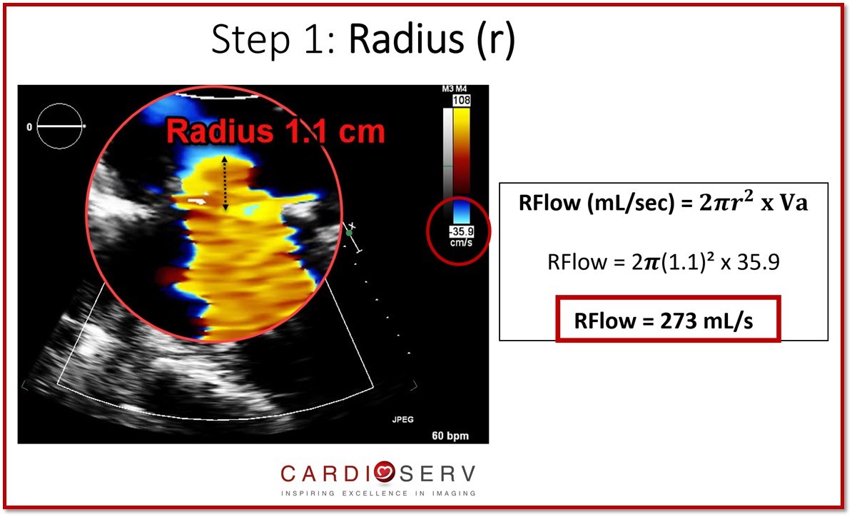
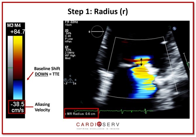
Step 1: Measure the Radius (r)
- Zoom on the Mitral Valve (MV) to optimize visualization of the regurgitant jet.
- Activate Color Doppler and adjust for a clear flow convergence hemisphere.
- Shift the color baseline downward (TTE) to around 20–40 cm/s to enhance aliasing.
- Freeze the image at mid-systole, when the MR jet reaches its peak.
- Measure the radius (r) from the aliasing boundary (first color change) to the vena contracta.
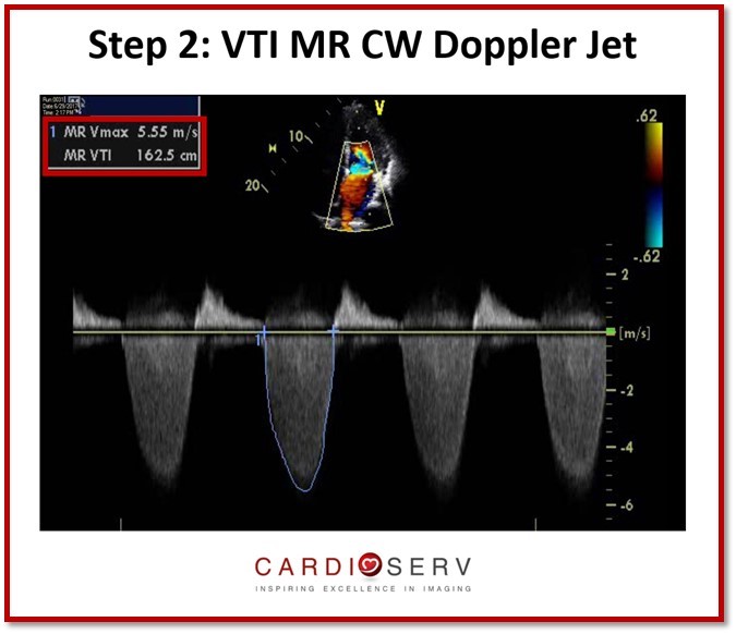
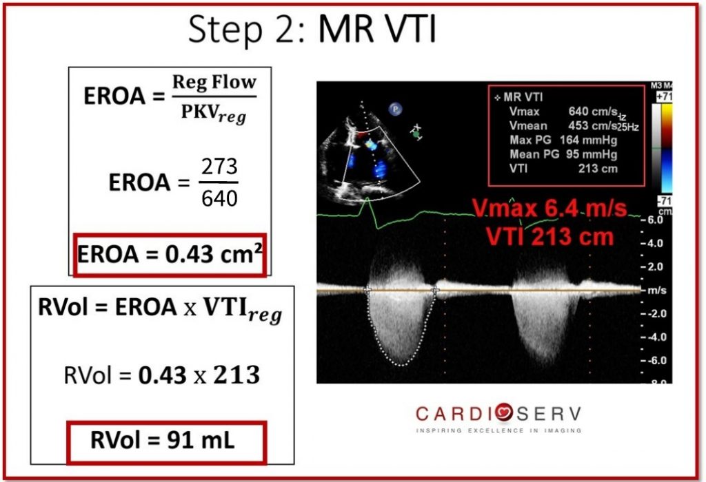
Step 2: Measure the MR Jet Velocity (CW Doppler)
- Zoom on the Mitral Valve and Left Atrium (MV/LA) for optimal alignment with the MR jet.
- Apply Color Doppler to identify the direction and origin of the regurgitant jet.
- Use Continuous-Wave (CW) Doppler to trace the MR jet velocity through systole.
- Measure the VTI (Velocity Time Integral) of the MR jet on the CW Doppler spectrum.
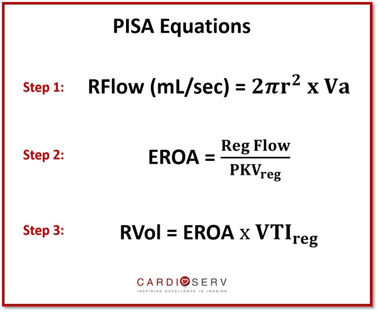
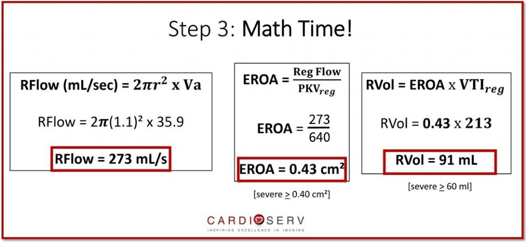
PISA Equations
Thankfully our machines do the math for us! But if you are the type of person that likes to know the breakdown process of how we obtain our outcome values, here are the equations!
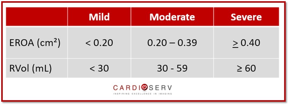
PISA Measurement Values
Use the following reference values to interpret the quantitative severity of mitral regurgitation using the PISA method.
Note: Always confirm MR severity using an integrative approach — combine PISA findings with other parameters such as vena contracta width, pulmonary vein flow, and LV/LA size and response (as emphasized by ASE Guidelines 2017).
PISA LIMITATIONS
- Timing matters: The CW Doppler and PISA radius must be obtained at the same point in the cardiac cycle—specifically, at mid-systole when flow convergence is largest.
- Single-frame calculation: PISA is derived from a single frame, assuming a hemispheric flow convergence that may not capture temporal variations in regurgitant flow.
- Non-holosystolic jets: PISA tends to overestimate MR severity when the regurgitant jet is not holosystolic. In such cases, regurgitant volume (RVol) may provide a more reliable estimate.
- Eccentric or multiple jets: Eccentric, wall-hugging, or multiple MR jets can distort flow convergence geometry, making accurate radius measurement difficult.
- Assumption of hemispheric geometry: The PISA method assumes a perfectly hemispherical flow convergence, which may not hold true in all cases, particularly with irregular or elliptical orifices.
Case Example
Now that we understand how to perform the PISA method, let’s walk through a case example!

Take the Guesswork Out of Mitral Valve Imaging
Mitral Valve Imaging in Echo – Visual Guide
Sharpen your MR assessment with this clear, image-rich guide to echo views, scallops, and probe positioning.
- ✔ Understand scallop anatomy in TTE & TEE with ease
- ✔ Avoid common pitfalls like foreshortened views
- ✔ Dozens of labeled diagrams for quick reference
- ✔ Practical checklists to streamline MR imaging

Andrea Fields MHA, RDCS
Stay Connected: LinkedIn, Facebook, Twitter, Instagram
References:
Zoghbi, W. A., MD, FASE, & Adams, D., RCS, RDCS, FASE. (2017). Recommendations for Noninvasive Evaluation of Native Valvular Regurgitation. JASE, 30, 4th ser., 1-69. Retrieved June 12, 2017. DOI: 10.1016/j.echo.2017.01.007