ASE’s Mitral Regurgitation Algorithm Simplified!
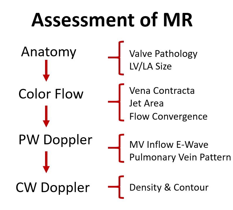
Now that we have covered all the methods for evaluating mitral regurgitation (MR), let’s put it to use! The ASE released an algorithm as a guide to help determine the severity of chronic MR, using both qualitative, semi-quantitative & quantitative measures.
ASE’s Mitral Regurgitation Algorithm Simplified! Read More »