August 2023

Understanding LVH Part 2: How to Measure LV Mass and Diagnose LVH

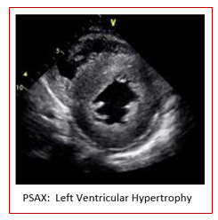
As diagnostic imaging professionals, we often perform echocardiograms on patients with hypertension to monitor the thickness, strength and wall motion of the heart. Last week we launched our two-part blog on left ventricular hypertrophy (LVH). In part one we explained the pathophysiology behind the various categories of LVH along with the echocardiographic findings. We discussed how LVM and RWT plays a role in identifying the categories of LVH. This week we will answer the question: What is LVM and RWT and how do we obtain these values?

Happy International Women’s Day!

As a company founded by and ran by women, CardioServ is proud to support International Women’s Day! A few years ago, our CEO and founder of CardioServ, Judith, went to Haiti to assess the diagnostic imaging needs of hospitals and clinics within Haiti for HCM. HCM serves over 80,000 people who would not usually have access to healthcare as well as organizing other education, nutrition and sustainability projects.

Understanding LVH Part 1: Concentric, Eccentric and Concentric Remodeling

Last month was designated American Heart Month to raise awareness of cardiovascular disease, the leading fatality of Americans. AHA encourages our patients to take control over understanding their risk factors of heart disease including knowing their numbers related to blood pressure. Untreated hypertension can have a long-term effect on the heart causing hypertensive heart disease, which can include coronary heart disease (CAD), left ventricular hypertrophy (LVH) and hypertrophic cardiomyopathy (HCM).

Helping Physicians “See”

For all of us involved in quality improvement measures within our lab our goal is to drive change. Reporting physician compliance with turnaround time or participating in group peer review sessions to track interpretive variability can often generate some lively conversations! As an educator and consultant, I have facilitated meetings where I have seen first hand interpretive variability or had to report on non-compliance with physician turnaround time. Take ejection fraction as an example of interpretive variability. Although ASE provides guidelines regarding best quantification techniques (Biplane Simpsons Method), the physicians are still faced with the reality of the sonographer’s ability to perform correct techniques while performing the exam! How do we close the loop on subjective ultrasound interpretation and tip the scales to consistently provide accurate interpretations? How do we understand the many issues involved with a physician that may not read a study on time?

Staff Spotlight: Andrea

We are starting our new series Staff Spotlight by introducing our newest employee Andrea Fields! Andrea is our new Cardiac Clinical Director. She will help develop our clinical and educational programs related to echocardiography. Her experience with advanced echocardiography will be a great addition to our team. Learn more about her below!

6 Steps to Meeting New IAC QI Measures

So over the past year we saw a lot of changes regarding the IAC accreditation quality improvement requirements across all modalities. Finally, as the dust settled we were left with a standardized program across the board. We expected the new QI to be difficult and time consuming so it took a while for us to get our head around just how simplified the whole system now is! We here at CardioServ are all loving the new workflow and we would like to share what we enjoy most about the new requirements.

Scroll to Top