Last week guest writers Dr. Ali and Dr. Nossikoff explained what an agitated saline “Echo Bubble study” is. They provided 7 indications for use of an echo bubble study along with a review of how to interpret the findings. If you missed last week’s blog, check it out! This week both doctors will teach us 9 steps to perform an echo bubble study along with supplies needed and tips to optimize imaging.
- Educator Spotlight: Dr. Ahmed Ali
- Educator Spotlight: Dr. Alexander Nossikoff
- 7 Indications for an Echo Bubble Study
ECHO BUBBLE STUDY RECAP
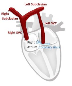
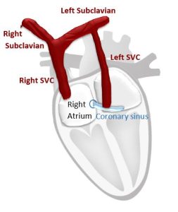
Agitated saline, or an echo “bubble study” is often a neglected complementary echocardiographic technique. An Echo Bubble Study is an injection of saline after agitation with air to create micro-bubbles that are ultrasound reflective into a vein in order to reach and opacify the right heart chambers, the coronary sinus in cases of persistent left superior vena cava (PLSVC), or the pericardium during pericardiocentesis.
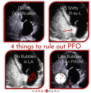
Review last week’s blog for a detailed explanation of indications for echo bubble studies and how to interpret the findings.
SUPPLIES NEEDED FOR AN ECHO BUBBLE STUDY.
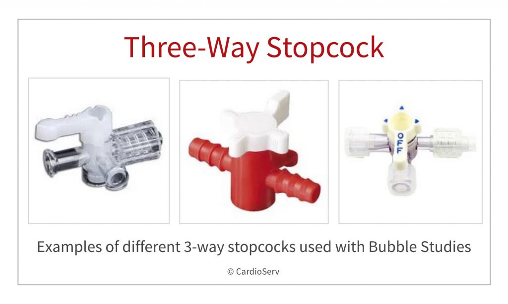
You will need:
1. IV Cannula
IV cannula, size is not so important as it is in contrast CT, forceful push immediately after agitation is far more important. However, larger cannula sizes (20 gauge or more) diminish the risk of vein rupture and make forceful injections easier. If you withdraw blood (see below) do it immediately after inserting the IV cannula directly into one of the syringes.
2. Three-way stopcock.
3. At least two 10ml syringes
At least two 10ml syringes (preferably 4-5, as you may need repeated injections and it is best practice to withdraw blood in the beginning) with locking mechanism (try to avoid regular syringes at they may leak during forceful injection).
4. Normal Saline 0.9 %.
5. Extension tube connector (optional).
6. Gelofusine (optional).
7. Assistance
2nd operator/nurse to help with injections (optional) – it is difficult to focus on both image acquisition and injection simultaneously
9 STEPS TO PERFORMING AN ECHO BUBBLE STUDY
Step 1:
1- Start by preparing all your required materials.
Step 2:
2- Fill one of the syringes with 8-9 ml of normal saline. Consider mixing saline with gelofusine in 50:50 manner, for example 4 ml or 4.5 ml of each, this is done routinely by many high volume centers.
Gelofusine strongly enhances contrast appearance, however this can lead to more distal shadowing. Keep in mind that gelofusine enhanced opacification stays much longer especially in cases of significant tricuspid regurgitation, which may cause problems when repeated injections are needed as you will have to wait much longer for complete clearance.
Step 3:
3- Insert IV cannula in an ante-cubital vein, in the most convenient arm for injection – if patient is lying on his left side use right arm and vice versa. However, if you want to show PLSVC use of left arm vein is mandatory.
Some operators use leg vein injections for PFO detection, as IVC flow directly hits the fossa ovalis region and may reveal shunts that are not visible with arm injection. This practice is very rare nowadays as the focus has shifted on maneuvers to increase right atrial pressure.
Step 4:
4 – Withdraw 0.5-1ml of blood in the syringe filled with saline as blood enhances contrast appearance. This is particularly important when not using gelofusine for enhancement. If gelofusine is used there is no value of adding blood as opacification is very strong anyway, so the blood withdrawal can be skipped.
Step 5:
5 – Connect the three-way stopcock to the cannula either directly or through an extension tube. The extension tube should be flushed with saline before connecting the syringe with blood to avoid spillage
Step 6:
6 – Withdraw 0.5 – 1 ml of air in the other syringe so you do not disconnect the one with blood in it. If no blood is withdrawn, there seems to be no advantage of withdrawing air in a separate syringe as contents would be mixed anyway during the forceful pushes, therefore you can withdraw the air in the syringe with the saline/gel already in it.
Step 7:
7- Start agitating the saline moving it forcefully between the two syringes connected to the three-way stopcock in a proper position so you do not lose the contrast too early in circulation during pushes. It is very important that the last push through the stopcock is forceful before injecting into the vein. Most experienced operators push backward and forward 3-4 times and then do one last very forceful push before injecting.
Step 8:
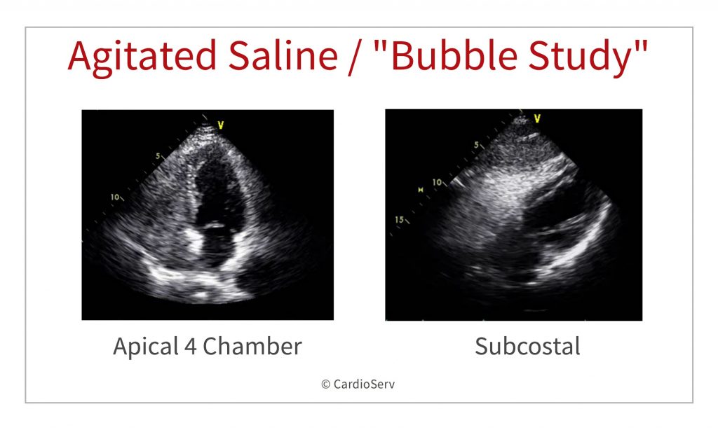
8- Once the required echocardiographic view is optimal; then rapidly and forcefully inject the agitated saline into the vein. Some operators prefer injecting with the syringe parallel to the vein (or to the extension tube) and then squeeze the forearm to enhance contrast entrance into venous system and respectively RA.
Step 9:
9- If the first injection (without Valsalva) is positive, no further images are necessary. If the first injection is negative, then the patient is asked to perform a Valsalva maneuver and agitated saline is injected before the patient releases it. Most operators consider that proper Valsalva is extremely important for PFO detection, the patient should strain appropriately for at least 10 seconds. Then Valsalva release causes a brief right-to-left shunt, because of increased pressure in right atrium, due to the rapid influx of blood from the IVC/SVC.
SAFETY OF AGITATED SALINE IN ECHO
Agitated saline injection is safe with very few (if any) side effects that are mostly related to injection site.
Traditionally, agitated saline use has been considered contraindicated if right-to-left shunting is known or suspected due to presumed risks of systemic air-embolism. However, it is well known that some degree of right-to-left-shunt is present in about 25-30% of healthy young population due to PFO. Actually, detection of PFO is the most common indication for doing agitated saline study. Data from large American databases shows that cerebro-vascular accidents caused by agitated saline injections used for PFO detection are extremely rare and frequently without long-term consequences. Use of gelofusine has been associated with rare cases of hypersensitivity reactions, but most experienced operators have never observed any so the rate should be very low.
A very simple safety tip for diminishing the amount of air entering the circulation can be used when performing agitated saline injections – bend the syringe tip down and the plunger up during injection (this is particularly easy when using extension tube), this way the air will stay up near the plunger, stop the injection when close to the last 2ml.
SUMMARY
In conclusion; agitated saline is a cheap, safe and easy to perform technique with lots of added information when properly used. Last week we reviewed the 7 Indications for a bubble study along with how to perform and interpret the findings. This week we reviewed how to perform an echo bubble study. Next week we will provide 6 tips to improve image quality when performing an echo bubble study. We would like to thank our guest writers this week.


Dr. Alexander Nossikoff
If you are interested in being a guest writer, please contact us


