This week we are kicking off the diastolic function guideline series, hosted by our guest blog writer Michael Owen! But first, we need to cover the basics on ‘how-to‘ acquire the measurements needed to evaluate for diastolic function.
The measurements needed to evaluate diastolic function include: pulsed-wave (PW) Doppler of mitral valve inflow, tissue Doppler imaging (TDI) and pulmonary veins, left atrial (LA) volume index, tricuspid regurgitation (TR) Doppler velocity and left ventricle (LV) ejection fraction (EF). Luckily, we have covered a few of these items in our past blogs!
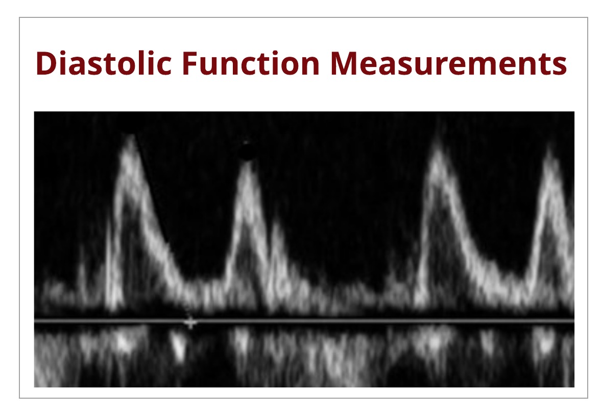
MITRAL VALVE INFLOW:
The first group of measurements come from performing PW Doppler at the mitral valve leaflet tips. The following measurements can be obtained from this waveform:
- Peak E-Wave Velocity (cm/s)
- Peak A-Wave Velocity (cm/s)
- A-Duration (msec)
- E/A Ratio
- Deceleration Time (msec)
HOW TO OBTAIN MITRAL VALVE INFLOW WAVEFORM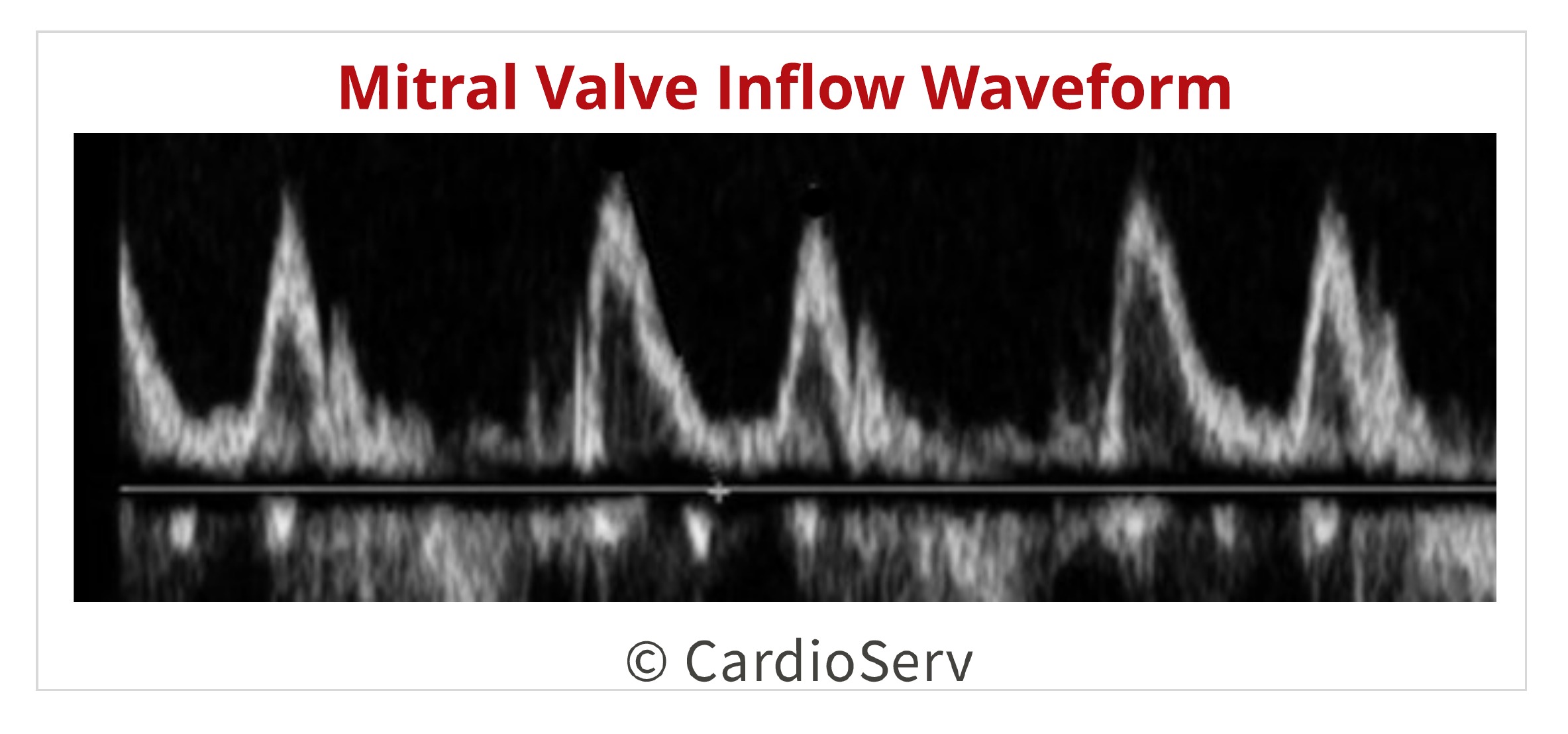
- Apical 4 chamber
- Pulsed-wave Doppler
- Sample volume between mitral valve leaflet tips
- Low wall-filter
MITRAL VALVE INFLOW MEASUREMENTS
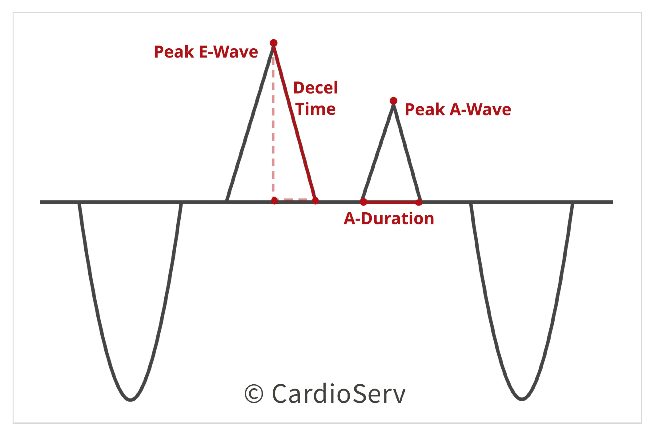
PEAK E-WAVE VELOCITY (CM/SEC)
- Peak modal velocity
- Early diastole (after T-wave on EKG)
- Leading edge of waveform
PEAK A-WAVE VELOCITY (CM/SEC)
- Peak modal velocity
- Late diastole (after P-wave on EKG)
- Leading edge of waveform
A-DURATION (MSEC)
- Time interval measurement
- Onset to end of A-wave along baseline
E/A RATIO
- E-wave velocity divided by A-wave velocity
DECELERATION TIME (MSEC)
- Time interval measurement
- Peak E-wave along slope of LV filing extrapolated to zero-velocity baseline
PW TISSUE DOPPLER IMAGING:
Next we need to evaluate the septal and lateral e’ velocity using PW tissue Doppler imaging (TDI)! Using this, we can obtain the following measurements:
- e’ Velocity septal/medial and lateral (cm/sec)
- Average E/e’
HOW TO OBTAIN PW TDI WAVEFORM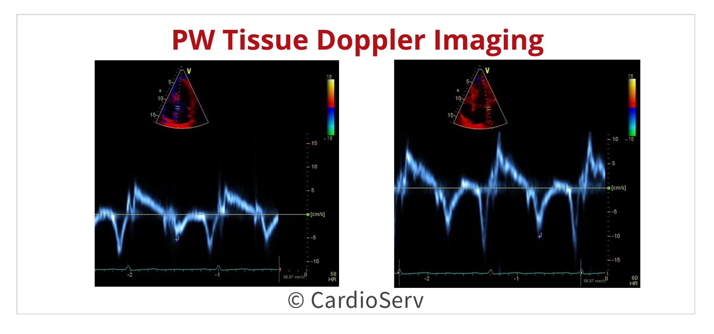
- Apical 4 chamber
- PW Doppler TDI
- Sample volume on septal and lateral basal regions
PW TISSUE DOPPLER IMAGING MEASUREMENTS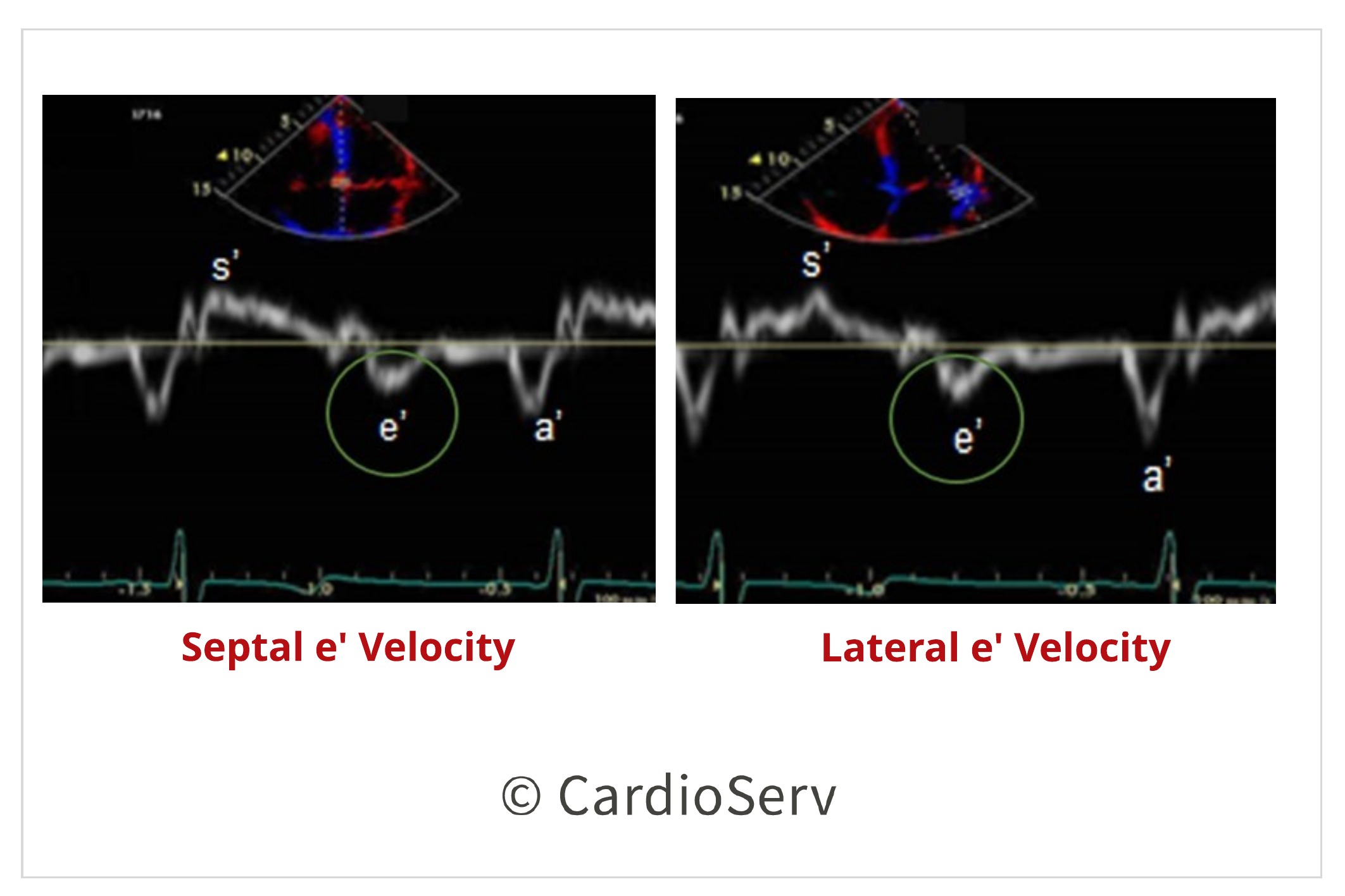
E’ VELOCITY (CM/SEC)
- Peak modal velocity
- Early diastole
- Leading edge of waveform
- Adjust gains for Doppler signal appears sharp
AVERAGE E/E’
- E-wave velocity divided by e’ velocity
PULMONARY VEINS
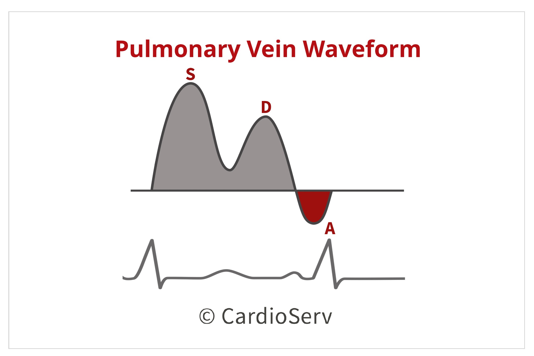
Lucky for you, we already posted a blog on correct tips to obtaining pulmonary vein Doppler waveform! You can review it here! The measurements needed from the pulmonary vein waveform for evaluation of diastolic function include:
- S-Wave (cm/sec)
- D-Wave (cm/sec)
- AR Duration (msec)
- S/D Ratio
PULMONARY VEIN MEASUREMENTS 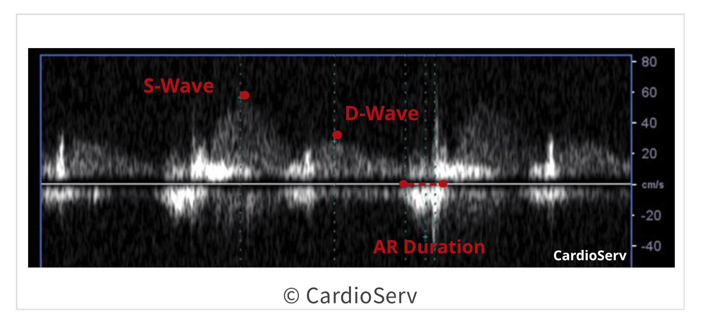
S-WAVE VELOCITY (CM/SEC)
- Peak modal velocity
- Early systole
- Leading edge of spectral waveform
D-WAVE VELOCITY (CM/SEC)
- Peak modal velocity
- Early diastole after MV opening
- Leading edge of spectral waveform
AR DURATION (MSEC)
- Time interval
- AR-wave onset to end of AR at zero baseline
S/D RATIO
- S-wave divided by D-wave velocity or
- S-wave VTI divided by D-wave VTI
LEFT ATRIUM SIZE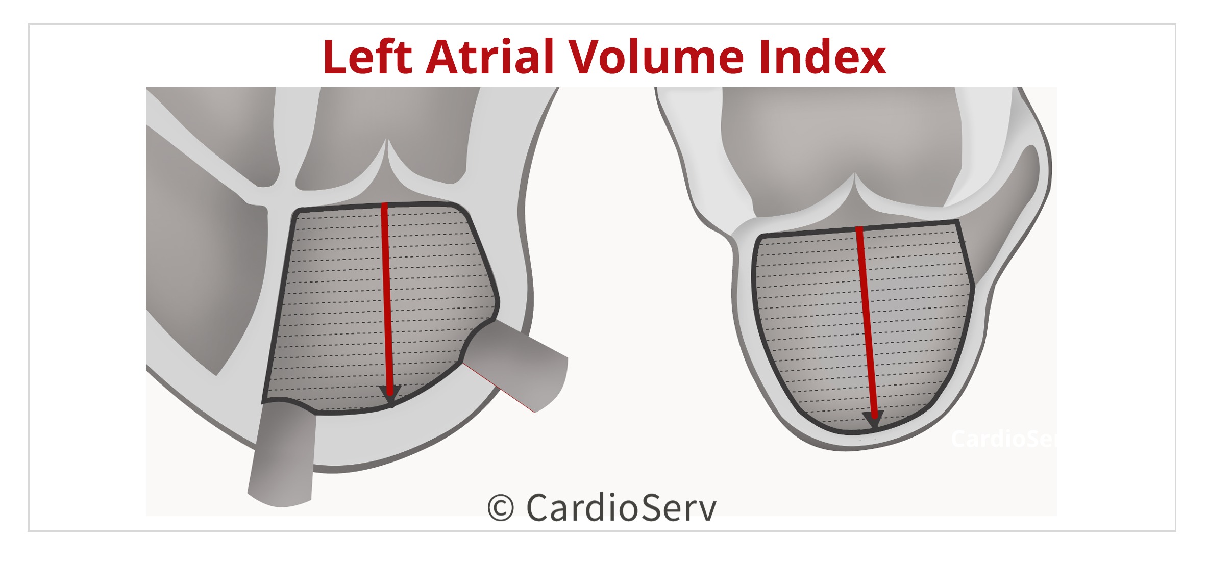
We also need to evaluate the left atrium volume, indexed to BSA (LAVI)! We have a blog written on this already- you can check it out here!
LEFT VENTRICLE EJECTION FRACTION 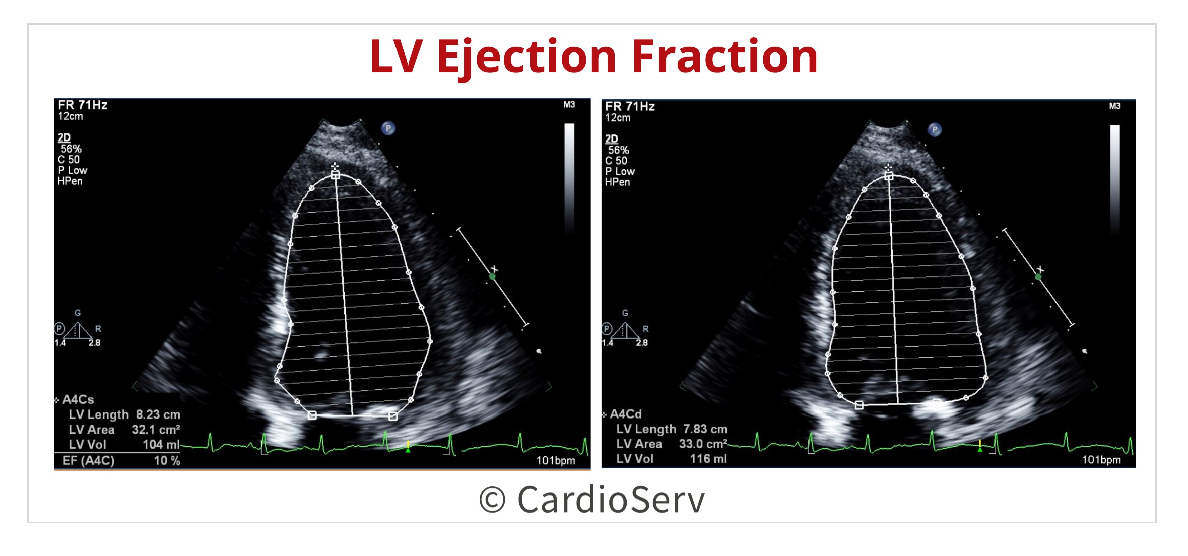
You can view the blog over proper method to performing LV bi-plane measurement to determine volumes and ejection fraction here!
TRICUSPID REGURGITATION 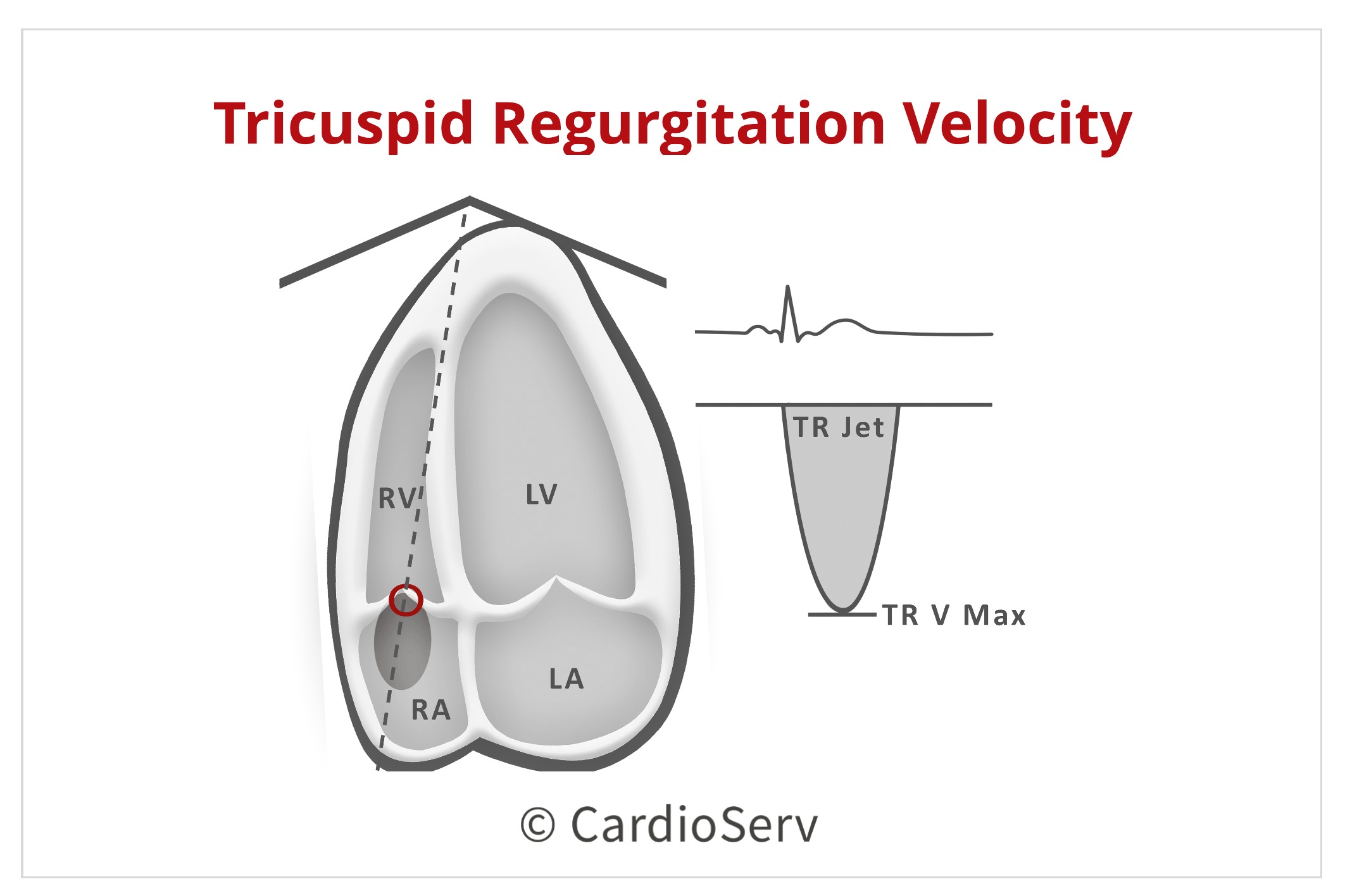
The last measurement we must have in order to evaluate for diastolic function is the tricuspid regurgitation (TR) velocity gradient! You can find out how-to guide from our right heart quantification blog series, here!
SUMMARY
Now that we have covered the basic assessment of measurements, we’re ready to dive into how to apply these towards determining diastolic function!

Andrea Fields MHA, RDCS
Stay Connected: LinkedIn, Facebook, Twitter, Instagram
References:
Nagueh, S. F., MD, Smiseth, O. A., MD, & Appleton, C. P., MD. (2016). Recommendations for the Evaluation of Left Ventricular Diastolic Function by Echocardiography: An Update from the American Society of Echocardiography and European Association of Cardiovascular Imaging. American Society of Echocardiography, 29(4), 277-314. Retrieved October 31, 2017, from http://asecho.org/wordpress/wp-content/uploads/2016/03/2016_LVDiastolicFunction.pdf