Last week we posted an article on acute pulmonary embolism in echocardiography. In that post we discussed how echo is good for ruling-in pulmonary embolism but not for ruling-out a pulmonary embolism. After diagnosing a pulmonary embolism what is the role of echo? Many studies have demonstrated the prognostic value of echo in determining short term mortality, prolonged hospital stay, and/or survival. This week we will explore TAPSE in the setting of a pulmonary embolism.
If you need to refresh your memory on your right heart quantification measurements, you can review past blog articles here:
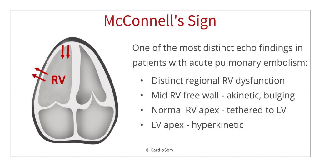
TAPSE IN MCCONNELL’S SIGN
First, let’s review TAPSE with McConnell’s sign. Last week we reviewed how the McConnell’s Sign is one echo finding in the setting of acute pulmonary embolism. The classic distinction of McConnell’s sign is normal RV apex wall motion (RV apex is tethered to the LV apex) and the mid free RV wall bulging out due to akinesis. Remember, the sudden pressure overload of the RV causes stress on the RV and this stress is not uniform causing localized mid free RV wall bulging.
DOES THE BASAL RV SEGMENT MOVE NORMALLY IN MCCONNELL’S SIGN?
One of our readers asked if TAPSE with McConnell’s Sign is normal. With the mid wall being akinetic, the basal wall may appear “normal” to the eye. Although, the basal wall moves better than the akinetic mid section of the RV – there is still dysfunction, therefore TAPSE would not be normal.
To avoid confusion, we’ve updated our image from last week’s blog by removing arrows from the basal RV segment.
Video from Daniel Migliaccio,
@TheScanimal Twitter Account,
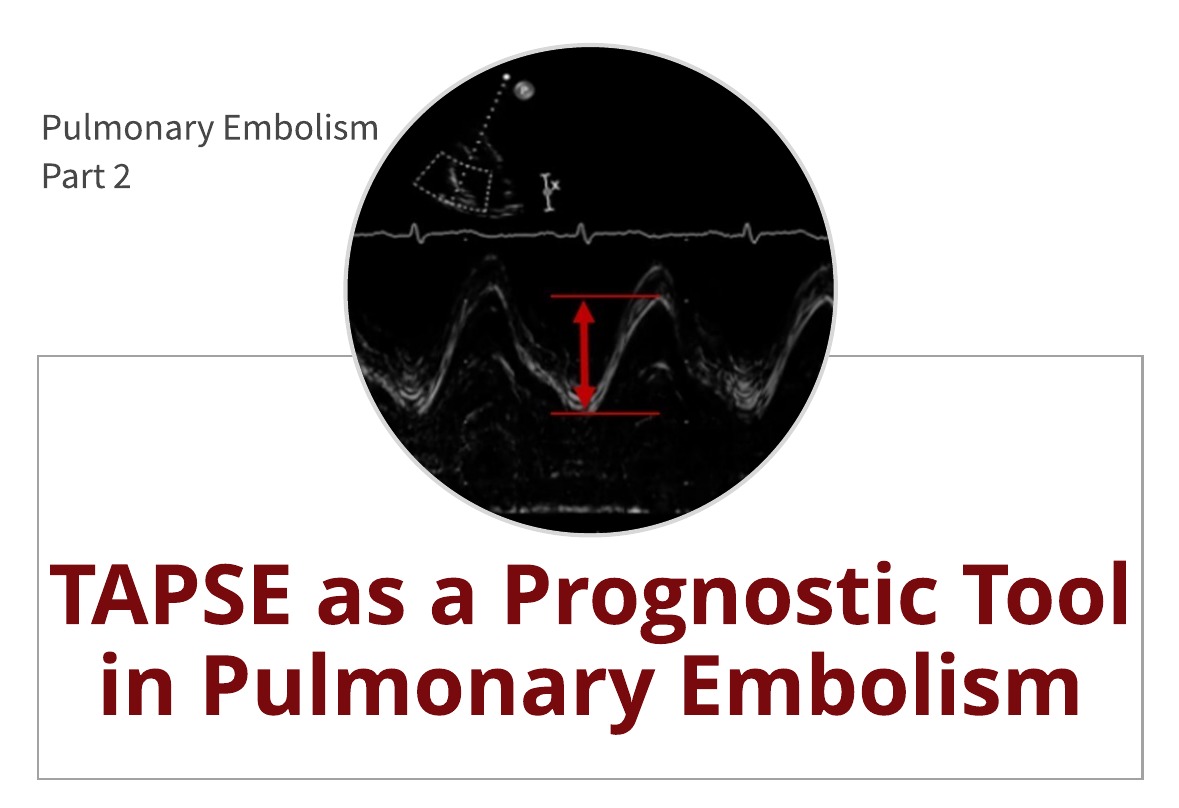
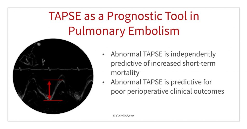
TAPSE AS A PROGNOSTIC TOOL IN PULMONARY EMBOLISM
So, how does echo help as a prognostic tool in pulmonary embolism? “Right ventricular failure remains a major cause of mortality during acute pulmonary embolism” (Schmid et al, 2015). Through the use of echo and quantification measurements like TAPSE, we have the ability to assess RV function. If we can identify patients with RV dysfunction, we can make better decisions on how best to treat them.
Read 8 Tips to Correct RV Function Assessment with TAPSE and S’ Wave for a step by step guide.
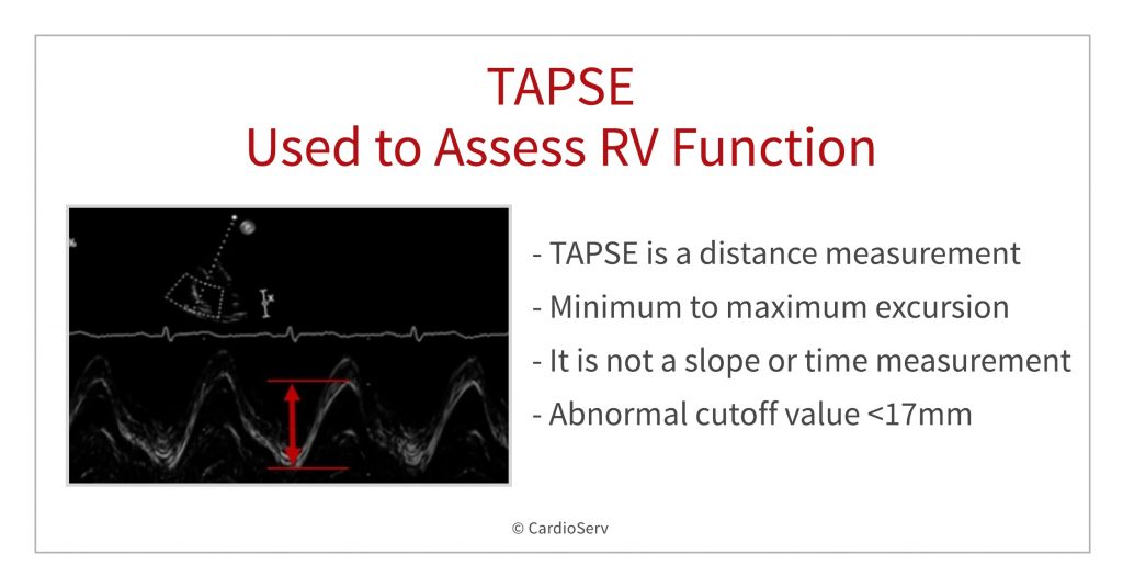
WHAT TAPSE TELLS US
- Decreased RV function and abnormal TAPSE is independently predictive of increase short-term mortality
- In a study with 1035 patients with PE, with preserved systemic artetial pressure, patients with RV hypokinesis and abnormal TAPSE had a 30-day mortality rate that was twice as high as patients with normal TAPSE
- Abnormal TAPSE is independently predictive of increased length of stay in ICU
- Abnormal TAPSE is predictive for poor perioperative clinical outcomes in the surgical setting of PE patients
- Abnormal TAPSE is an independent risk factor for intraoperative CPR and death.
- Study with 81 patients undergoing pulmonary embolectomy of the 11 patients that required intraoperative CPR (11/81) all had abnormal TAPSE
- Study with 81 patients undergoing pulmonary embolectomy of the 8 patients that died intraoperatively all had abnormal TAPSE
SUMMARY
Right ventricular hypokinesia and abnormal TAPSE are independent predictors of increased risk (double) for 30-day mortality in patients with acute pulmonary embolism. Abnormal TAPSE is also predictive of poor perioperative outcomes. TAPSE is easy to perform and is reproducible between users and readers. Please review how to correctly perform TAPSE.
CAT 1. AMA CMES – RIGHT HEART QUANTIFICATION
If you are in need of CMEs, please take advantage of our right heart quantification bundle package. All CMEs are online and offer printable certificates upon completion with unlimited access to educational material. In addition, each course includes a free eBook!
References
- Butts, C., MD. (2017, March). The Speed of Sound: A New Measure to Single Out High-Risk PE Patients: Emergency Medicine News. Retrieved from https://journals.lww.com/em-news/Fulltext/2017/03000/The_Speed_of_Sound__A_New_Measure_to_Single_Out.7.aspx
- Hickey, S., MD, & Alerhand, S., MD. (2018, August 27). Tricuspid Annular Plane Systolic Excursion (TAPSE) for Risk Stratifying Patients with Pulmonary Embolism. Retrieved from http://www.emdocs.net/tricuspid-annular-plane-systolic-excursion-tapse-for-risk-stratifying-patients-with-pulmonary-embolism/
- Lobo, J.L., et al., Prognostic significance of tricuspid annular displacement in normotensive patients with acute symptomatic pulmonary embolism.J Thromb Haemost, 2014. 12(7): p. 1020-7.
- Prats, M. (2017, August 28). TAPSE to Diagnose Pulmonary Embolism. Ultrasound GEL. Retrieved from https://www.ultrasoundgel.org/posts/EJHu_SYvE4oBT4igNHGBrg
- Rydman, R., et al., Echocardiographic evaluation of right ventricular function in patients with acute pulmonary embolism: a study using tricuspid annular motion.Echocardiography, 2010. 27(3): p. 286-93.
- Vitarelli, A., et al., Right ventricular function in acute pulmonary embolism: a combined assessment by three-dimensional and speckle-tracking echocardiography.J Am Soc Echocardiogr, 2014. 27(3): p. 329-38.