This week we are going to cover our last of the three quantification methods for evaluating the severity of mitral regurgitation (MR). The past two weeks we discussed PISA and stroke volume methods. If you missed them or would like to refresh, you can find them there:
VOLUMETRIC METHOD
The Volumetric Method is very similar to the Stroke Volume method. It is based on the same principles as the stroke volume method. The difference is that with the volumetric method, the SV across the MV inflow is replaced with the Bi-Plane Simpson’s measurements. Let’s explore this step by step.
First lets refresh the basic concept behind both the Stroke Volume Method and the Volumetric Method.
- When blood is pushed out of the LV during systole, some of the regurgitant blood is pushed back across the mitral valve.
- The stroke volume at both inflow and outflow sites are obtained to calculate the amount of regurgitant volume across a leaky valve.
- The stroke volume for inflow can be calculated either using:
- SV across the MV using the MV diameter width and PW Doppler, as discussed last week, or
- Bi-plane Simposon Method which we will discuss this week.
The volumetric method provides us with:
- Stroke Volume (SV) of the LV
- Regurgitant Volume (RVol)
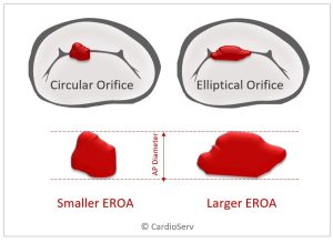
- Effective Reguritant Orifice Area (EROA)
HOW DO WE CALCULATE THE SV OF THE LEFT VENTRICLE?
Last week we discussed how to calculate SV of both inflow (mitral valve) and outflow (aortic valve) using the cross sectional area formulas. If you need a refresher, you can find it here! This week we will continue to use the same method for calculating the stroke volume across the aortic valve but review a more common method for calculating the LV volume.
The recommended method to obtaining volumes of the left ventricle, is by the Simpson’s Biplane measurement in both apical 4 and 2 chambers. Review our blog on best practices for performing LV volume via Biplane Simpson, here.
HOW TO PERFORM VOLUMETRIC METHOD
It will be necessary to calculate the following two values:
- SV of LV
- SV of LVOT (Aortic Valve)
STEP 1: SV OF LV (SIMPSON’S BIPLANE)
- Apical 4 and Apical 2 Window
- Trace LV at end-diastole
- Trace LV at end-systole

STEP 2: SV OF OUTFLOW (AORTIC VALVE)
- Parasternal Long Axis (PLAX) Window
- LVOT Diameter
- Apical 5 Window
- PW LVOT Velocity- VTI

STEP 3: CW DOPPLER MR JET VELOCITY & MEASURE
- Zoom MV/LA
- Color Doppler
- CW Doppler MR
- VTI MR Velocity

VOLUMETRIC METHOD EQUATIONS
If you like to perform the math without the ultrasound machine help, here are the equations you’ll need:

VOLUMETRIC METHOD VALUES

VOLUMETRIC METHOD LIMITATIONS
- Underestimating true LV volumes
- Avoid foreshortening
- Use contrast for suboptimal images
- Easy to underestimate regurgitant severity
- Do not use m-mode for volumes
CASE EXAMPLE
Let’s walk through a case example utilizing this method!


SUMMARY
This week wraps up our last method to quantify the severity of mitral regurgitation! If you’ve missed our blog series covering everything you need to know in regards to echo and MR, you’re welcome to check them out here! We hope you continue to enjoy reading our blogs and able to take away good information to implement into your department!

Andrea Fields MHA, RDCS
Stay Connected: LinkedIn, Facebook, Twitter, Instagram
References:
Zoghbi, W. A., MD, FASE, & Adams, D., RCS, RDCS, FASE. (2017). Recommendations for Noninvasive Evaluation of Native Valvular Regurgitation. JASE, 30, 4th ser., 1-69. Retrieved June 12, 2017.


