Last week we discussed 1 of the 3 ways to quantify the severity of mitral regurgitation (MR), using the PISA method (proximal isovelocity surface area). If you missed it, you can find it here! This week, we are going to explain the second method– stroke volume method & provide a case example on how it’s performed! Our goal is to help you easily understand the concept and process of implementing the stroke volume method for evaluation of MR into your echo lab!
STROKE VOLUME METHOD
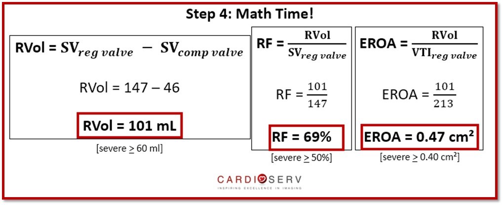
This method is based off of the regurgitant volume (RVol) calculation: Difference between the inflow and outflow stroke volumes (SV) of the left ventricle (LV).
Concept based on: if there is no regurgitation present, the stroke volume at both sites are equal.
The SV Method provides us with:
- Regurgitant Volume (RVol)
- Regurgitant Fraction (RF)
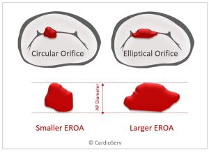
- Effective Regurgitant Orifice Area (EROA)
WHAT IS STROKE VOLUME
Stroke Volume is the amount of blood (volume) pumped by the heart with each beat. In a heart with normally functioning valves, absent of regurgitation, the volume entering the left ventricle across the mitral valve (inflow) will be equal to the volume exiting the left ventricle across the aortic valve (outflow). Meaning, the SV across the aortic valve equals the SV across the mitral valve.
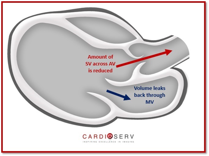
WHAT HAPPENS IN THE PRESENCE OF MITRAL REGURGITATION?
As the heart pumps blood out of the left ventricle during systole, some of the volume leaks back through the regurgitant mitral valve. When this happens, the SV across the aortic valve (outflow) is reduced (as some of the volume is escaping through the leaky MV). This difference in volume is how the regurgitant volume is calculated. Lets review how to calculate the inflow and outflow stroke volumes and learn how to use this information to calculate the regurgitant volume.
HOW DO WE CALCULATE SV?
The two valves we will be focusing on to determine the SV method for MR are the Mitral Valve (inflow) and the Aortic Valve (outflow).
You can calculate the SV of an orifice by obtaining two measures:
- Cross-sectional Area (CSA)
- Inflow VTI at the Annulus

It’s important to know that we must obtain the measurement for both values at the same location. We calculate the SV for each valve during their normal flow period:
- Mitral Valve: Diastole
- Aortic Valve: Systole
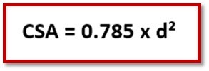
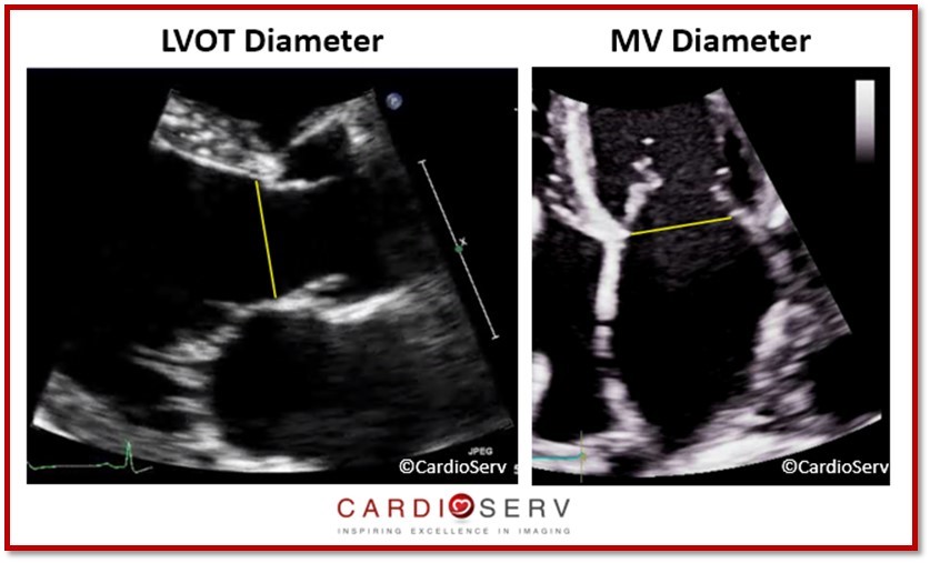
CROSS-SECTIONAL AREA (CSA)
This method assumes the annulus is circular. We can calculate the CSA of an annulus by obtaining the diameter at the location.
- Mitral Valve: Early-Diastole & Inner-edge to Inner-edge
- Aortic Valve (LVOT Diameter): Mid-Systole & Inner-edge to Inner-edge
INFLOW VTI OF ANNULUS
For each annulus, we measure the pulsed-wave (PW) Doppler at the normal flow pattern of the valve.
- Mitral Valve (Diastole)– above the baseline
- Aortic Valve [LVOT] (Systole)– below the baseline
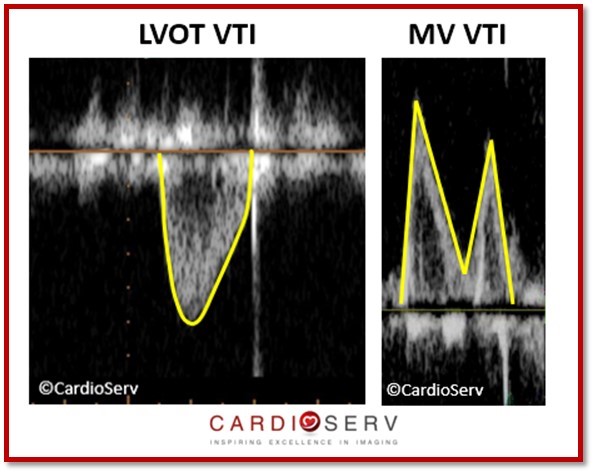
**REMEMBER: Place the sample volume at the SAME location as diameter was taken. This is vital for the MV because we are use to placing the sample volume at the leaflet tips to evaluate diastolic function–however: for determining MV SV, we must place the sample volume gate at the MV annulus!
HOW TO PERFORM SV METHOD
In order to obtain the values for this quantification method, we must follow these steps:
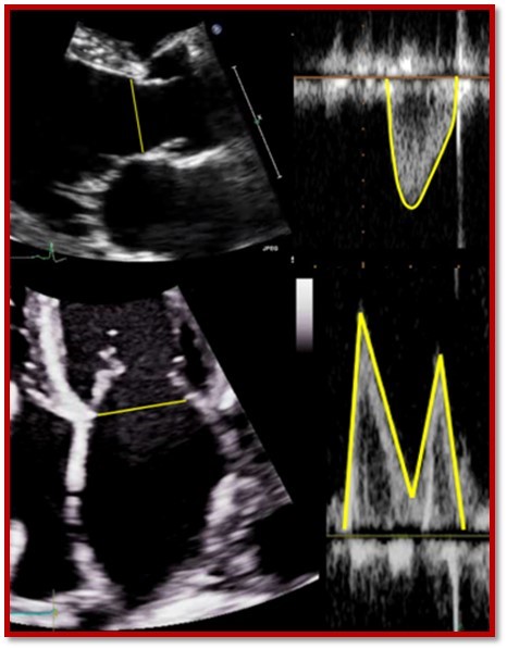
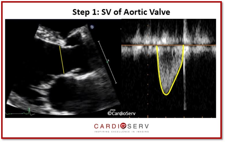
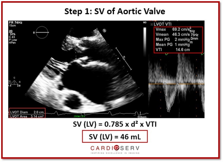
STEP 1: MEASURE SV AORTIC VALVE OUTFLOW
- Parasternal Long Axis (PLAX): LVOT Diameter (mid-systole)
- Apical 5 Window
- PW Doppler at LVOT
- VTI Outflow Velocity
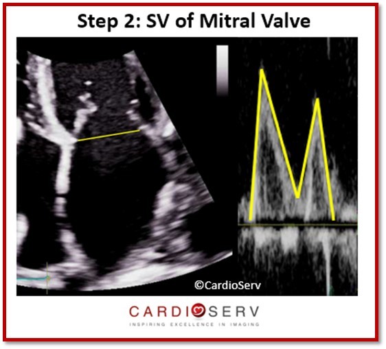
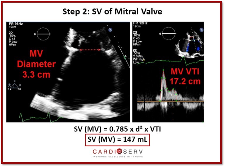
 STEP 2: MEASURE SV MITRAL VALVE INFLOW
- Apical 4 Window
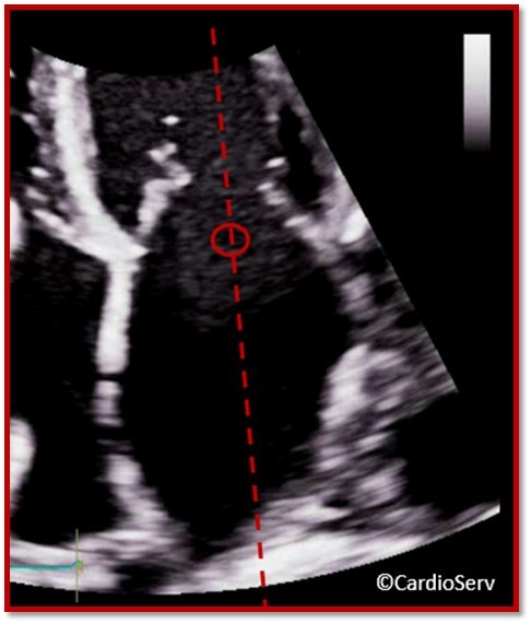
- MV Diameter Width @ Annulus (early-diastole)
- PW Doppler at Annulus
- VTI Inflow Pattern
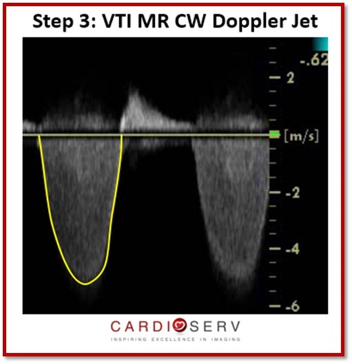
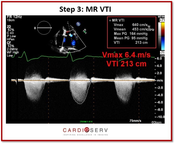
STEP 3: CW DOPPLER MR JET VELOCITY & MEASURE
- Zoom MV/LA
- Color Doppler
- CW Doppler MR
- VTI MR Velocity
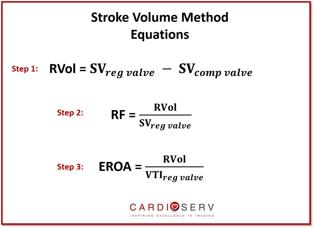
STROKE VOLUME EQUATIONS
Your ultrasound machine package should calculate this for you. Just remember to correctly use your measurement package to identify each measurement that you make. For example, if you just place generic calipers on a frozen image and measure the MV annulus, this value will not be entered into your calculation package. You must first select MV annulus within y0ur measurement package for the system to assign this value and correctly calculate your formula. If you like to perform the math on your own, here are the equations to do so:
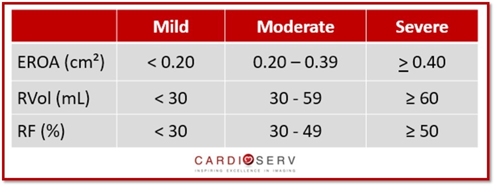
STROKE VOLUME METHOD VALUES
STROKE VOLUME LIMITATIONS
- Incorrect annulus measurement
- Incorrect velocity tracing
- Incorrect sample volume placement
CASE EXAMPLE
Now that we’ve discussed how to perform this method of quantification, let’s break down a case example!
SUMMARY
This week we have added another method to quantifying the severity of MR. We hope you can add this method, along with the PISA method we discussed last week. Keep an eye out for the 3rd way, next week, over the volumetric method.
We appreciate all of our readers and love to hear feedback! Be sure to check out our other blogs on the website!

Andrea Fields MHA, RDCS
Stay Connected: LinkedIn, Facebook, Twitter, Instagram
References:
Zoghbi, W. A., MD, FASE, & Adams, D., RCS, RDCS, FASE. (2017). Recommendations for Noninvasive Evaluation of Native Valvular Regurgitation. JASE, 30, 4th ser., 1-69. Retrieved June 12, 2017.