
Last Updated on October 17, 2025 by Don Gerig, RDCS
In Hepatic Veins 101, we discussed the structure, function and waveforms of the hepatic veins, in regards to the cardiac cycle. This blog will cover how the hepatic veins play a role with the right heart. We tend to forget about the importance and useful information the hepatic veins provide us. Lets review, how the hepatic veins play a role in:
- Tricuspid regurgitation (TR)
- Right heart failure
Tricuspid Regurgitation
The hepatic vein waveform can be reflective on the severity of TR present. The more severe the regurgitation, the more pulsatile the waveform becomes.
Waveform Criteria:
- Pulsatile waveform
- Tall A-wave & V-wave
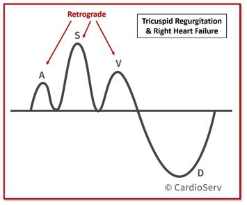
- Decreased or Reversed S-Wave
- D-Wave < S-Wave
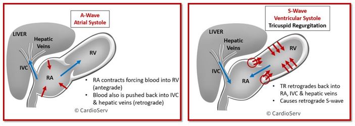
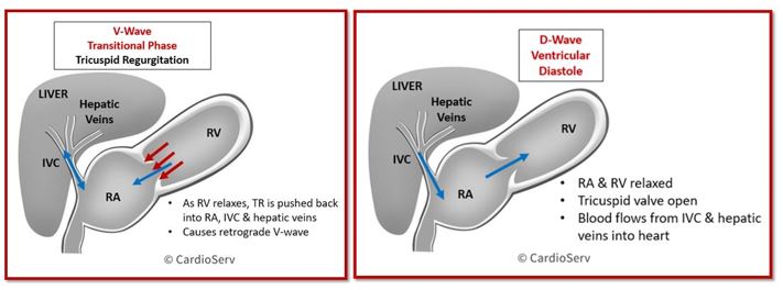
Let’s breakdown the waveform process during the cardiac cycle when tricuspid regurgitation is present….
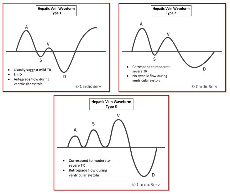
A scale was developed based upon the waveform pattern of the hepatic veins to help with adding more data to assess the severity of TR:
Right Heart Failure
The hepatic vein waveform can also show indications of right heart failure! The change in waveforms are similar to tricuspid regurgitation…
Right Heart Failure Criteria:
- Pulsatile waveform
- Tall A-wave & V-wave
- Normal relationship between S-wave & D-wave (S > D)
- S-wave will never become retrograde (in absence of TR)

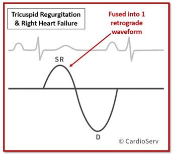
Combined Tricuspid Regurgitation and Right Heart Failure
In some cases, where the patient has both TR and right heart failure, the hepatic waveforms can become biphasic.
- Single retrograde wave per cardiac cycle
- Single antegrade wave per cardiac cycle
In severe cases, the waveforms can fused into one retrograde wave with an antegrade D-wave.
Items to Note:
- Hepatic vein waveforms look the same for both Mild TR and for right heart failure (both can have type 1 or 2 waveform patterns)
- Retrograde S-wave present = some degree of TR is present
Hepatic Vein Systolic Filling Fraction
The Systolic Filling Fraction (SFF) is the ratio of systolic forward flow (S-wave VTI) to the total forward flow (S + D-wave VTI), excluding the A-wave:
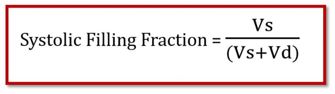
A normal SFF indicates that most venous return occurs during systole, when right atrial pressure is low. As RAP increases, systolic flow decreases, lowering the SFF.
An SFF < 55% correlates with an RAP > 8 mmHg, showing 90% sensitivity and specificity — even in ventilated patients when averaged over multiple beats.
The higher the RAP, the lower the Systolic Filling Fraction.
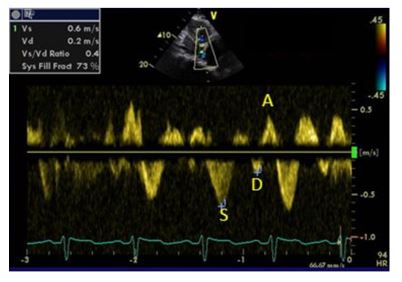
VS/VD Hepatic Vein Ratio
By measuring the velocity of the S-wave and D-wave, we can obtain a simple ratio to help indicate if an elevated RAP is present.
Vs/Vd < 1 = Elevated RAP
- Higher the right atrial pressure (RAP), the lower the pressure gradient (PG) between hepatic veins/IVC and right atrium
- S-wave diminishes due to decrease of systolic flow
- D-Wave (Vd) Dominant
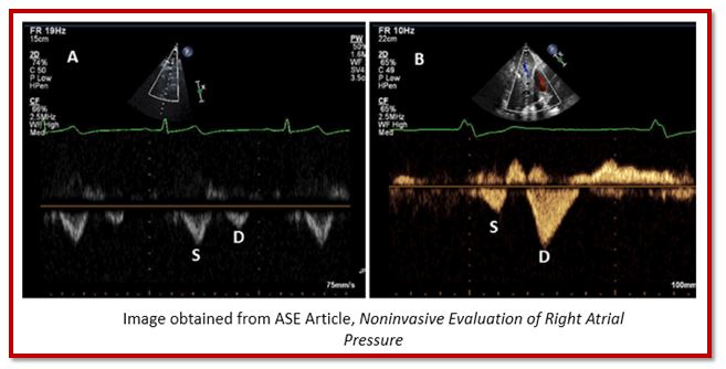
Let’s take a look at this image below:
- On the left, a normal waveform pattern is demonstrated showing S/D >1
- One the right, an abnormal waveform pattern is demonstrated showing S/D <1
- Indication of elevated RAP
Summary
Now that we have learned both the basics of hepatic veins and the relativity in regards to the heart, we can now implement evaluation of these veins when indicated during our echocardiograms! Here are a few helpful tips to take away…..
- Hepatic veins are useful for determining RAP when the IVC is unable to assess for collapsibility
- Hepatic vein waveform can help determine severity of TR and presence of right heart failure
- Pulsed Doppler the hepatic vein at close proximity to IVC
- Dilated hepatic veins (>10mm) can indicate right heart failure and TR
- Hepatic veins can be useful in diagnoising additional pathology, in additional to TR and right heart failure
- If >moderate TR present, evaluate the hepatic veins
We hope you enjoyed our right heart blog series!
Go Deeper
Ready to strengthen your understanding right-heart hemodynamics?
Enroll in our Right Heart Quantification & Hemodynamics CME course for a step-by-step review of the 2025 ASE guideline framework. You’ll learn how to integrate Doppler, IVC, and TR data for accurate pressure estimation—and earn CME credit while doing it.

Stay Connected: LinkedIn, Facebook, Twitter, Instagram
References:
Scheinfield, M. H., MD, Bilali, A., ARDMS, & Koenigsberg, M., MD. (2009). Understanding the Spectral Doppler Waveform of the Hepatic Veins in Health and Disease. RadioGraphics,29, 2081-2098. Retrieved May 31, 2017, from http://pubs.rsna.org/doi/pdf/10.1148/rg.297095715
Rudski, L. G., MD. (2010). Guidelines for Echocardiographic Assessment of the Right Heart in Adults: A Report from the American Society of Echocardiography. JASE, 23, 685-713.


