We love it when our readers post comments and ask questions. We respond to every question. A couple of weeks ago we were asked about the assessment of RVSP in the presence of wide open TR with poor TV coaptation. What a great question! We will answer that this week. Please keep your questions coming!
ETIOLOGY OF TR
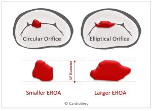
Most TR is functional rather than pathologic (75%) and is accompanied by lengthening of the TV leaflets in an effort to maximize the coverage of the TV annulus to prevent backflow. However, regardless of the cause for non-coaptation of the TV leaflets, severe TR reflects the equalization of RV and RA pressures. In this setting, the PASP (which is equal to RVSP in the absence of pulmonic valve stenosis) may be inaccurate (overestimated)
PASP AND RAP
In the estimation of PASP, we have the added dilemma of estimation of the right atrial pressure (RAP). Fortunately, the mild and severe ranges of pulmonary hypertension are easier to estimate accurately. The middle or moderate range proves harder to narrow down. According to the ASE article Echocardiography in Pulmonary Arterial Hypertension: from Diagnosis to Prognosis, a peak systolic velocity of <2.8 m/sec is usually present in the absence of pulmonary hypertension. Other values which can help with the diagnosis of pulmonary hypertension are given in this ASE article.
OTHER ECHOCARDIOGRAPHIC SIGNS
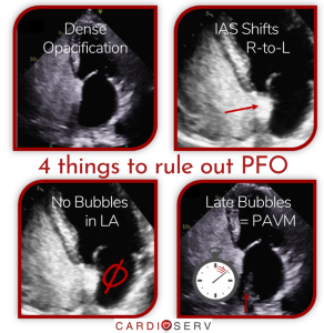
Whenever other echocardiographic signs are encountered, one must give consideration as to their contributing effect on the estimation of any Doppler-derived calculation. For instance, RV structure and function are important when assessing for pulmonary hypertension. Consider the differences between RV volume and pressure overload. Volume overload is “handled” by the RV better than is pressure overload. Of course, volume and pressure overload can exist together. Echocardiographic signs such as a D-shaped septum (one which does not round up during diastole is indicative of RV pressure overload) and an interatrial septum which bows to the left are strong indicators of pulmonary hypertension.
OTHER TECHNIQUES
Other techniques such as TDI and strain can help in evaluating RV function. Three dimensional imaging has the distinct advantage of allowing for better correlation with cardiac MRI as far as RV volumes are concerned.
CLOSING THOUGHTS
Echocardiography serves as an important means of diagnosing and classifying pulmonary hypertension. As such, we, as cardiac sonographers, owe it to our patients to use all the tools at our disposal correctly. In order to do our best, we must stay on top of the current best practices and guidelines by craving knowledge. Thank you for your thought provoking question. I hope that these comments and articles help!

Yvonne Prince ACS, RDCS, RVT, RDMS, FASE
Connect on LinkedIn!
RELATED ARTICLES:
- 8 Things to Know About Estimation of RAP via Echocardiography
- 2 Ways to Properly Assess TR Jets for Accurate RVSP Calculations
- What the Heck is the Cut-Off Value for RVSP?!
- 6 Tips for Calculating RVSP
- TAPSE and RVSP- Prognostic Value When Viewed as an Index
References
http://asecho.org/wordpress/wp-content/uploads/2014/09/33-2013-J-Am-Soc-Echocardiogr-Echocardiography-in-Pulmonary-Arterial-Hypertension.pdf.


