MITRAL REGURGITATION
It is estimated that over 4 million people in the United States have significant mitral regurgitation. Untreated severe mitral regurgitation decreases a person’s quality of life and has a high mortality rate. With the advancement of technology, including the MitraClip, more procedures and treatment options are available. The accurate quantification of mitral regurgitation plays a critical role in determining the best course of action for patients living with MR. This week we will review how to measure the MR Vena Contracta Width.
First of all, its important to remember that the process of MR quantification is an integrated process that includes:
- Color Doppler
- PW Doppler
- CW Doppler
- Advanced Quantification
In this blog, we will cover vena contracta width (VCW). The vena contracta width is just one measure derived from color Doppler. A complete review of MR Quantification can be found on our learning platform cme.cardioserv.
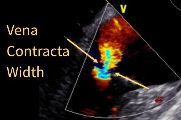
MR VENA CONTRACTA WIDTH (VCW)
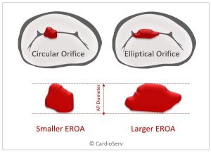
The Vena Contracta is the narrowest portion of the MR jet and in the presence of a circular-shaped MR orifice, the cross-sectional area directly represents the orifice area which is a true parameter of MR severity.
MR Vena Contracta Width: Pros
- Semi-quantitative measure
- A good marker to distinguish mild from severe
- Can be used for both central and eccentric jets
- Independent of driving pressures (blood pressure, etc.) for a fixed MR orifice – Eg. Rheumatic heart, annular calcification, endocarditis, etc.
MR Vena Contracta Width: Cons
- Small measurement errors lead to misclassification of MR severity
- MR NOT holosystolic (MVP) = overestimation
- Multiple jets = underestimation
- Elliptical-shape orifice (secondary MR) = underestimation
MR VCW DIAGNOSTIC CRITERIA
The VCW values that are relevant to Doppler Quantification of mitral regurgitation are the high and low ends of the spectrum. Any value that is less than 0.3 cm is an indicator of mild MR and any VCW value that is equal to or greater than 0.7 cm is an indicator of severe MR.
The values in-between do not represent moderate but rather indicates a need for additional quantification.

MR VENA CONTRACTA WIDTH: STEP-BY-STEP INSTRUCTIONS
- Parasternal long-axis view
- Ultrasound beam perpendicular to flow
- Adjust the focus setting to the level of the MV
- Zoom image
- Color Doppler sector – NARROW around the valve (maximize lateral & temporal resolution)
- Color scale 50-70 cm/sec
- Scroll frame by frame through the systolic cycle for the largest jet area
- Measure the vena contracta – the narrowest portion of the jet
- Toggle the color on and off to better visualize the orifice
Tip: Must be able to see the flow convergence zone for an accurate measurement!
MR Vena Contracta Width (VCW) Summary
The Vena Contracta Width is a semi-quantitative measure for the assessment of MR severity. It is one measurement towards the bigger picture of an integrated approach to MR quantification. For a complete review of MR Quantification, see our 5 credit CME course: Mastering Advanced MR Quantification
Other Blogs you may like:
- Mitral Valve Anatomy: Name 5 Components!
- Smart Strategies for Determining MR Mechanisms
- Ultimate Guide to Acute vs. Chronic MR
- Secondary MR: Evaluating Mitral Valve Tethering
- FYI: Temporal Variation & Blood Pressure Matters for MR!
- 7 Factors that Influence Color Doppler Appearance
References:
American Society of Echocardiography. Recommendations for Noninvasive Evaluation of Native Valvular Regurgitation