Persistent Left Superior Vena Cava (PLSVC) creates a connection from the SVC to the coronary sinus. It is something we do not routinely see on echo but is always a great catch when we do see it! This week we will review the subclavian anatomy and blood flow of a PLSVC.
PERSISTENT LEFT SUPERIOR VENA CAVA (PLSVC)
PLSVC is an isolated anomaly with minimal hemodynamic and clinical significance. That said, there is a significant association with rhythm disturbances and other congenital cardiovascular abnormalities.
- Often discovered following the finding of an abnormally positioned catheter, pacemaker, or internal defibrillator lead
- If central venous catheter’s tip is in the left paramediastinal region – think PLSVC
- Confirm the presence of a PLSVC to rule out catheter perforation or migration
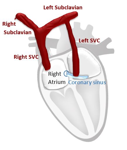
SUBCLAVIAN ANATOMY
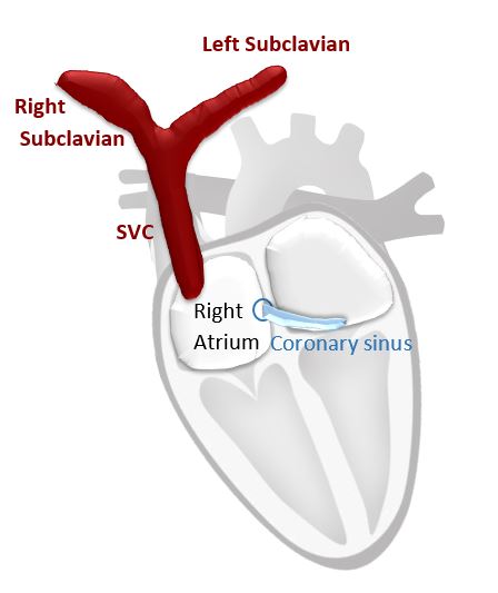
Normal Anatomy
- Right subclavian and left subclavian merge into the Superior Vena Cava (SVC)
- Normal flow – Superior Vena Cava to Right Atrium
Abnormal Anatomy
- Various forms of persistent left superior vena cava exist, all with the left and right subclavians separating and draining into a left and right superior vena cava. The left superior vena cava (LSVC) drains directly into the coronary sinus.
- PLSVC flow = Left superior vena cava into the VC to the coronary sinus

PLSVC Blood Flow
Blood flows from the left SVC into the coronary sinus and into the right atrium.

SUMMARY
This week we reviewed the anatomy and blood flow of a persistent left superior vena cava (PLSVC). Next week we will review how this presents during an echo bubble study.
Other articles you may like:
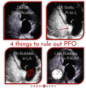
- 4 Things Needed To Rule Out a Patent Foramen Ovale (PFO)
- 7 Indications for an Echo Bubble Study
- 9 Steps to Perform an Echo Bubble Study
- 6 Tips to Improve an Echo Bubble Study!
EARN 2.5 CMES FROM OUR ONLINE COURSE
2.5 CMEs The Why and How of Echo Bubble Studies
- Preparation and Administration of Agitated Saline
- Tips for Improving Imaging
- PFOs
- Persistent Left Superior Vena Cava
- Pulmonary Arteriovenous Malformation
- Guided Procedures and Bubble Studies