ASE guidelines

Essential Steps to Evaluating MR Etiologies

The ASE recently released their new 2017 valvular regurgitation updated guidelines back in April. The paper details the difference between primary and secondary mitral regurgitation (MR) and outlines proper evaluation methods for the most common pathologies seen. The past couple of weeks, we have discussed how to differentiate between primary vs. organic and acute vs. chronic. If you need to refresh or missed our blog, you can read them here:

Finally… Mitral Valve Orientation Explained!

Last week, we covered the basic anatomy of the mitral apparatus. We reviewed in detail, the various structures and their function. As we continue with our mitral regurgitation blog series, we want to touch next on, specific imaging windows to evaluate the mitral valve. Remember, when we examine the valve, we need to make sure we include all structures of the apparatus.

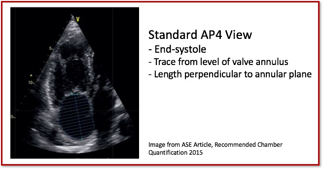
5 Things to Know about Using Fractional Area Change (FAC) to Assess RV Function

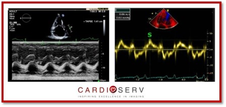
Before jumping into assessing the RV with the Fractional Area Change method, lets recap the past couple weeks. Last week, we talked about two methods for quantifying the function of the RV (TAPSE & S’ Wave). We selected these methods to explain first due to ease, reproducibility and established prognostic value. As some of our readers pointed out and as we discussed the TAPSE and S’ Wave methods do have limitations though. The 3 main limitations are:

10 Tips for Correct RA Size Quantification

We have been discussing the importance of evaluating the right heart over the past few blogs this month. The ASE updated the chamber quantification guidelines that provided us with the correct methods to measuring size and function of the right ventricle (RV) and atrium (RA). This week, we are going to talk about the proper method to measuring the size of the right atrium! We will review both the preferred volumetric measurement methods and the linear measurements of the right atrium.

Scroll to Top