Diastolic Function

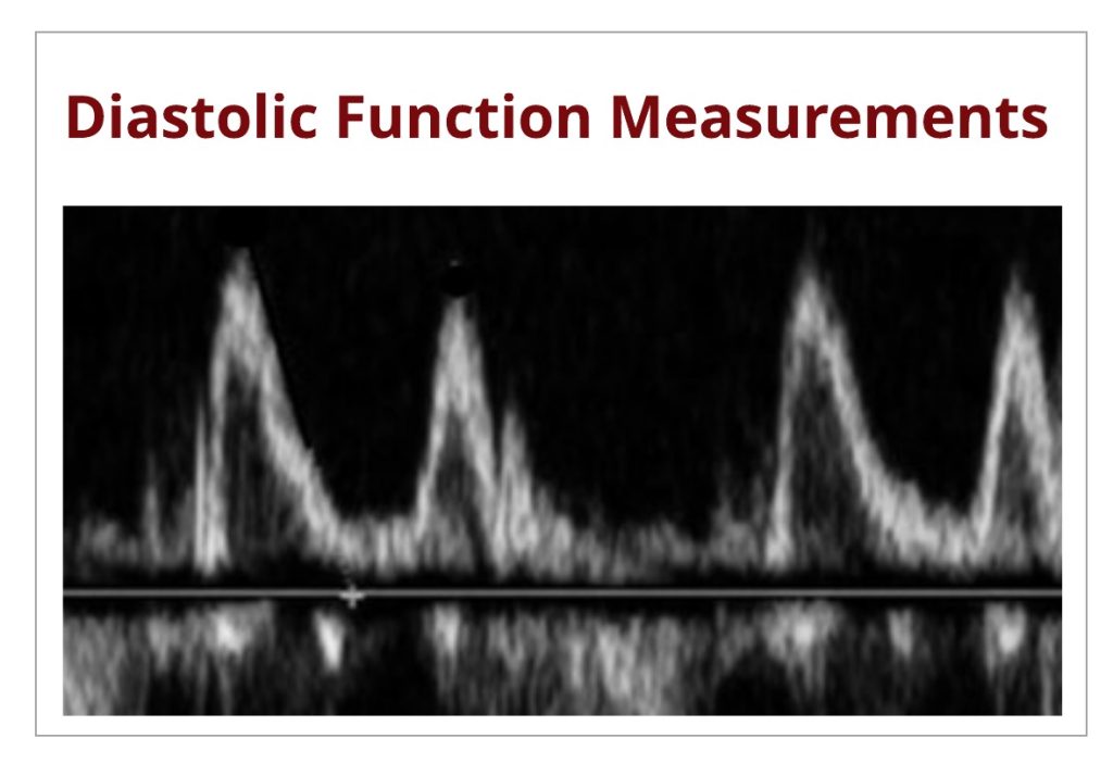
Avoid These Diastology Measurement Errors

Now that diastology is a mandated part of the scanning protocol for all of our accreditation clients, we are starting to see more diastology measurement errors. We thought that sharing these errors may help others to avoid them. This week, we will review correct tissue Doppler measurement techniques while reviewing two common errors. These errors include:

Understanding the Basics: Evaluating Diastolic Function

Diastolic function is a complex process that simply refers to how well the heart performs during diastole, as blood is suctioned from the left atrium (LA) towards the apex of left ventricle (LV) at a low-pressure filling state. When the heart is unable to fill properly, filling pressures increase in order to allow proper filling and keep required cardiac output state. When these filling pressures increase, diastolic dysfunction occurs.

Understanding the Basics: Physiology of Diastole

We are breaking down the basics of diastolic dysfunction into easy to understand blogs! Last week we discussed the topic of diastole and the 4 stages that occur during this phase within the cardiac cycle. If you missed it, you can read it here! This week, we are going to go a step further and discuss the physiology in regards to the 4 stages of diastole. We are going to cover:

Understanding Diastole

In the past weeks we had guest writer, Michael Owen, share how to assess diastolic dysfunction. He broke down the algorithm that ASE uses to evaluate the presence of dysfunction. The full ASE article is long and intense. Michael did a great job at simplifying the evaluation process. While featuring this Mastering Diastology series we started to receive a lot of questions. We realized that it would be helpful to take a step back and review the basics. All this great information regarding diastolic dysfunction means nothing to us unless we have a basic understanding of the topic and how it relates to our daily use. We are going to go back to the beginning. Let’s talk diastole!

Mastering Diastology: Part 2

Last week we discussed the algorithm for determining the presence of diastolic function in patients with normal ejection fraction (EF). This week, we are going to discuss determining the presence of diastolic function in patients with depressed EF or pathologic left ventricular hypertrophy (LVH) with preserved EF.

Mastering Diastology: Part 1

This week starts off our diastolic function blog series with our guest blog writer, Michael Owen! We introduced our followers to him last week- if you missed it, you can find it here! Our goal for this blog is to have a basic understanding of the normal ejection fraction (EF) algorithm for evaluating diastolic function!

Industry Spotlight: EchoToday

CardioServ loves promoting organizations and individuals that share our values of inspiring excellence. I interviewed EchoToday’s founder Michael Owen to find out more about his development of an app to advance workflow within imaging labs. Michael has mastered diastolic function evaluation and has found a way to simplify diastolic assessment with an app, allowing sonographers and physicians to quickly evaluate diastolic function.

Scroll to Top