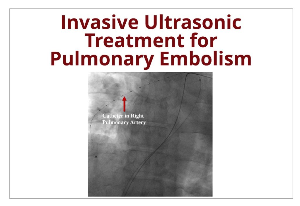
Invasive Ultrasonic Procedure to Treat Pulmonary Embolisms
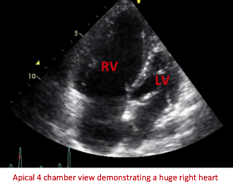
Pulmonary embolism (PE) is a critical condition where one or more of the arteries within the lungs become occluded by a blood clot. Quick diagnosis and treatment is crucial to keep mortality rates low. It is common for PE’s to originate from lower limb deep venous thrombosis (DVT). In other words:
Invasive Ultrasonic Procedure to Treat Pulmonary Embolisms Read More »