Written by Angela Mills, BA, RVS
What’s wrong with this vascular image?
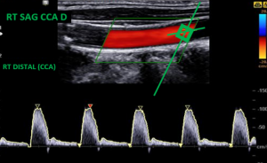
Modality: Carotid Duplex
View: Long axis CCA
Answer:
This image demonstrates prox CCA that was labeled and measured as a distal CCA.
Correct Technique:
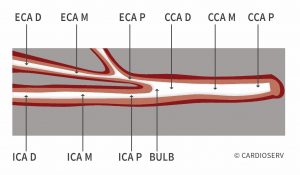
- The Proximal CCA placement should be very low on the neck usually close to the collar bone
- The Distal should be no more than 1-2 cm from the bifurcation.
LET US KNOW WHAT YOU THINK...


Feb
2017
Feb
2017
Feb
2017
Feb
2017
Feb
2017